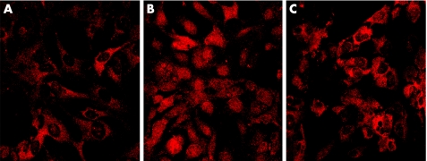
Figure 3 Immunocytochemical analysis of NF‐κB p65 localisation in HLE B‐3 cells after treatment with H2O2 in the absence or presence of PDTC. A: In untreated cells, NF‐κB p65 primarily existed in the cytoplasm, not in the nuclei. B: After stimulated with 0.5 mM of H2O2 for 1 hour, NF‐κB p65 translocated from the cytoplasm into the nuclei. C: In the cells pretreated with 100 μM of PDTC for 2 hours, few NF‐κB p65 translocated into the nucleus.

An official website of the United States government
Here's how you know
Official websites use .gov
A
.gov website belongs to an official
government organization in the United States.
Secure .gov websites use HTTPS
A lock (
) or https:// means you've safely
connected to the .gov website. Share sensitive
information only on official, secure websites.
