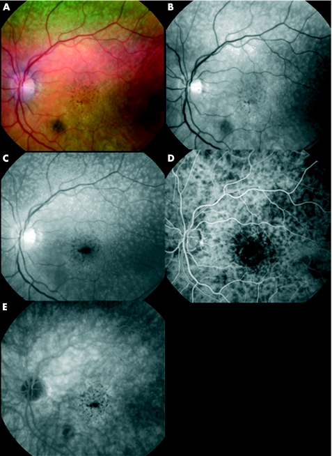
Figure 1 Reticular pseudodrusen (RPD). Colour (A), red‐free (B) and blue filter (C) fundus photographs; early (D) and late (E) frames of the fluorescein angiography of a patient with age‐related maculopathy characterised by macular pigmentary changes. RPD are barely visible on the colour picture (A), more clearly visible on the red‐free picture (B) and clear on the blue filter picture (C), on which they form a lacy pattern located outside the macula, near the temporal vessels. On fluorescein angiography, a patchy choroidal filling is present (D), but RPD are silent (D,E).

An official website of the United States government
Here's how you know
Official websites use .gov
A
.gov website belongs to an official
government organization in the United States.
Secure .gov websites use HTTPS
A lock (
) or https:// means you've safely
connected to the .gov website. Share sensitive
information only on official, secure websites.
