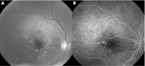
Figure 4 Reticular pseudodrusen (RPD) associated with retinal choroidal anastomosis or retinal angiomatous proliferation (RAP). Blue filter fundus photograph (A) and fluorescein angiogram (B). RPD are visible in the upper part of the macula (A). On the fluorescein angiogram (B), the temporal haemorrhage (A) corresponds to RAP, located in the temporal part of the macula (arrow).

An official website of the United States government
Here's how you know
Official websites use .gov
A
.gov website belongs to an official
government organization in the United States.
Secure .gov websites use HTTPS
A lock (
) or https:// means you've safely
connected to the .gov website. Share sensitive
information only on official, secure websites.
