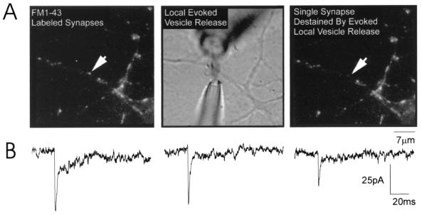
Figure 1.
Stimulation of mEPSCs at single synapses on cultured hippocampal neurons. (A) (Left) An image of the synapses on a portion of a dendrite of a hippocampal neuron maintained in vitro for 9 days. Each synapse is labeled with FM1–43; the arrow points to the single synapse from which mEPSCs were recorded. Before local perfusion, the synapse was clearly stained with FM1–43 and there was no electrical activity recorded because CNQX was present in the bath. Local perfusion was established by placing electrodes on either side of the selected synapse (Center; see Methods). Hypertonic solution containing no CNQX was puffed onto the synapse for 10 sec, every 10 sec; within milliseconds, mEPSCs were recorded. The stream was approximately 2 μm wide and never reached the neighboring synapse. The final image (Right), showing that the perfused synapse destained whereas neighboring synapses lost no fluorescence, provided confirmation that recorded mEPSCs originated exclusively from the selected synapse (arrow). (B) Representative dual-component mEPSCs recorded from a putative single synapse (cell 1 in Table 1).

