
Figure 3.
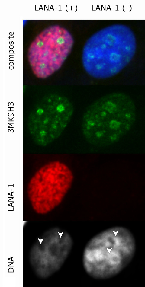
Dissolution of DNA from perinucleolar heterochromatin is not accompanied by the release of trimethylated lysine 9 histone H3 (3MK9H3) – green immunofluorescence staining in LANA-1 transfected cells (red). 3MK9H3 staining clearly identifies perinucleolar areas (white arrows) with diminished heterochromatic DNA staining in the transfected cells.