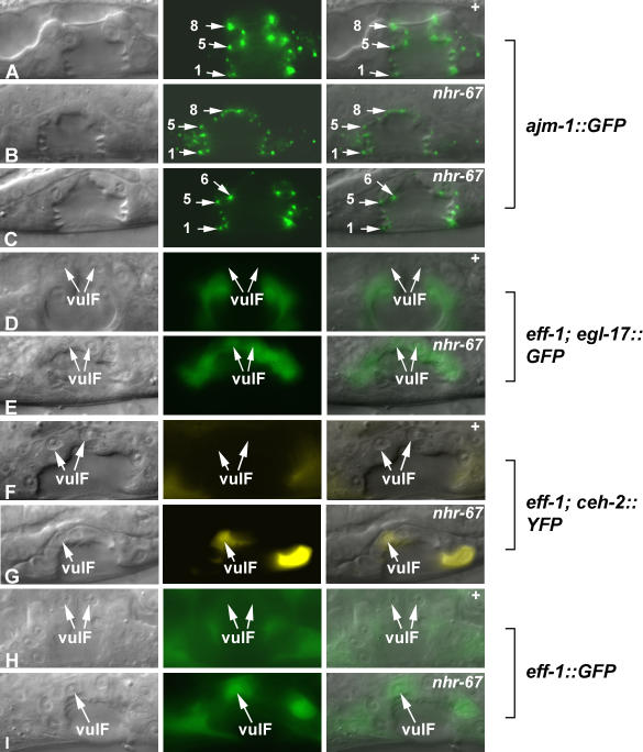
Figure 2. nhr-67 Prevents Inappropriate Fusion Events between the 1° Vulval Cells.
(A–I) Nomarski (left), fluorescence (center), and overlaid (right). The adherens junction marker ajm-1::GFP is used to visualize the cell number and architecture of the vulval toroids in wild-type (A) and nhr-67 RNAi–treated animals (B and C). When observing a mid-sagittal optical section of L4 hermaphrodites, ajm-1::GFP appears as dots between the vulval cells. Loss of adherens junction expression signifies a reduction in the cell number due to a cell fusion defect. (A) In wild-type animals, the eight dots on either side correspond to the seven distinct vulval cell types (arrows). The overall vulval morphology of nhr-67 RNAi–treated animals appears abnormal compared to wild-type.
(B) In some cases, the number of adherens junctions is normal in nhr-67 RNAi–treated animals (arrows).
(C) Reduction of nhr-67 sometimes results in the loss of dots at the top of the vulval invagination (which indicates an inappropriate fusion event between the vulE and vulF cells) (arrows). However, the altered gene expression observed in an nhr-67 RNAi background does not appear to be dependent on cell fusion defects.
(D) In the absence of eff-1-mediated fusion, ayIs4 [egl-17::GFP] expression is completely absent in the vulF cells (arrows).
(E) In contrast, depletion of nhr-67 activity in an eff-1 mutant background is sufficient to cause derepression of egl-17 in the 1° vulF cells (arrows).
(F) In eff-1 mutants, ceh-2 expression is absent in vulF cells (arrows).
(G) Reduction of nhr-67 activity in an eff-1 mutant background results in ectopic ceh-2 expression in vulF cells (arrow).
(H) In wild-type animals, eff-1::GFP is not expressed in vulF cells (arrows).
(I) eff-1 levels are elevated in vulF cells when nhr-67 activity is compromised (arrow).

