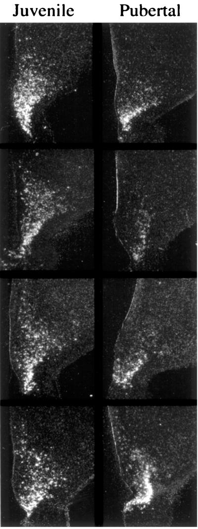
Figure 6.
Dark-field photomicrographs of emulsion-dipped hemicoronal sections (25 μm) taken through the midtuberal region of the MBH of four juvenile (Left) and four pubertal (Right) monkeys and hybridized with a 33P-labeled hNPY riboprobe. The ependymal lining of the third ventricle is visible on the left of each section. NPY mRNA expressing neurons are observed in the region of the arcuate nucleus in all animals. Labeling in areas dorsal to the arcuate region was more pronounced in juveniles. Labeling was absent in sections hybridized with sense riboprobe (not shown). (×8.)

