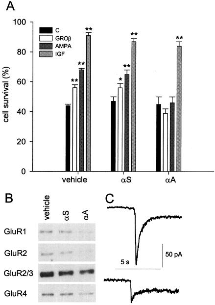
Figure 4.
Effects of antisense treatment on granule cells. (A) Cultured granule neurons were incubated with vehicle, αS, or αA as described in Material and Methods, and then were not treated (C), or treated with 60 nM GROβ, 10 μM AMPA, or 25 ng/ml insulin-like growth factor (IGF) for 24 h. Data expressed as in Fig. 1. Student's t test: ∗, P < 0.05; ∗∗, P < 0.001. (B) Western blot analysis of the expression of GluR subunits in cultures treated with vehicle, αS or αA. Band intensity quantification for the experiment shown (representative of four different determinations) revealed the following values (expressed as percentage of untreated cultures): GluR1: αS, 79%; αA, 9.5%; GluR2: αS, 63%; αA, 3%; GluR2/3: αS, 73%; αA, 56%; GluR4: αS, 83%; αA 30%. (C) Average currents from AMPA-stimulated nerve cells (30 μM; n = 10) untreated (Upper) or treated with αA (Lower) were, respectively 158 ± 39 pA (SEM, Student's t test, P < 0.05) and 60 ± 14 pA (SEM, Student's t test P < 0.05). Note larger reduction of AMPA currents in C compared with the GluR subunits expression in αA-treated neurons in B, which is attributed to the selection of most viable (likely antisense-underloaded) cells in C.

