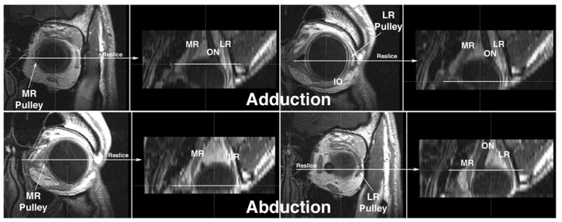
Fig. 2.

Gadodiamide enhanced, multiplanar T1 MRI of left orbit of normal human in adduction and abduction. Images were originally acquired in 18 contiguous 2 mm thick quasi-coronal planes perpendicular to the long axis of the orbit at 312 micron resolution. Using three-dimensional technique, the image stacks for each gaze direction were then interactively reconstructed in the planes indicated to produce the axial images shown. The horizontal rectus pulleys are recognized as dark rings In each case, the coronal images that include the medial rectus (MR) and lateral rectus (LR) pulleys are directly displayed for each gaze direction, and the corresponding location on the axial reconstruction is marked with a horizontal white line. Note that the MR pulley shifts posteriorly in adduction, and anteriorly in abduction. The LR pulley shifts anteriorly in adduction, and posteriorly in abduction.
