
Figure 1. LIN-17::GFP was asymmetrically localized within the B cell in lin-17 males prior to and after cell division.
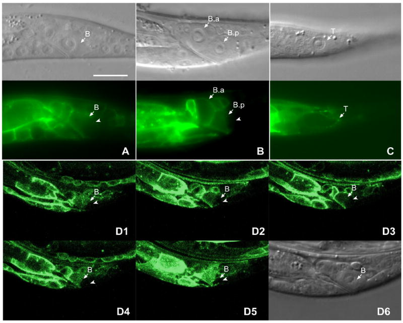
Panels (A–C) show the DIC image above and corresponding fluorescent image below. LIN-17::GFP was asymmetrically localized within the B cell prior to (A) and after division (B). The posterior cortex and cytoplasm (arrow) of the B cell (A) or the B.p cell (B) displayed lower level of GFP intensity than the anterior of that cell. (C) LIN-17::GFP was asymmetrically localized to the T cell and accumulated at the posterior cortex (arrowhead) of T cell. (D1–D5) show fluorescence confocal images of 1 μm Z sections through the B cell spatially sectioned from top to bottom. (D6) is the DIC image of B cell shown in D1–D5. Bar equals 10 μm in all panels.