
Figure 2. LIN-44 and MIG-5 were required for LIN-17::GFP asymmetric localization.
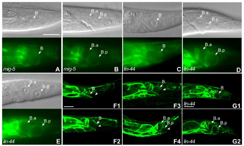
Panels (A–E) show the DIC image above and corresponding fluorescent image below. LIN-17::GFP was symmetrically localized within the B cell in mig-5 males (A) and lin-44 males (C), (compare to Fig. 1A). After the B cell division, the asymmetric localization of LIN-17::GFP was lost in mig-5 (B) and either reversed (D) or lost in lin-44 males (E) (compare to Fig. 1B). (F1-F4) localization of LIN-17::GFP at different time points during the B cell division: five hours after hatching (F1), before the B cell was about to divide (F2), during the B cell division (F3) and after the B cell division (F4). Arrowheads in F1-F4 point to the cell membrane or cortex that displayed lower GFP levels in the B or B.p cells. Bar in (A) equals 10 μm for (A–E) and bars in (F1) and (G1) equal 10 μm for (F1–F4) and (G1–G2).