
Figure 4. Cdk7 function is required for mitotic entry.
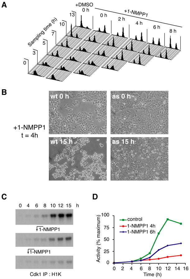
(A) Execution point determination of a Cdk7-dependent function in S/G2. Cdk7as/as cells were arrested at the G1/S boundary by double-thymidine block, then released into fresh medium without or with 10 μM 1-NMPP1 added at the indicated times. To monitor cell cycle progression, we measured DNA content at indicated times by flow cytometry. Note: after release from the thymidine block, we consistently observed a minor population of cells that remained arrested with a G1 DNA content (evident, for example, in the profiles of the DMSO control culture at 3, 5 and 7 h). (B) Inhibition of Cdk7 at 4 h after release from double-thymidine block prevents mitotic entry. Wild-type (wt) and mutant (as) cells were released into nocodazole-containing medium; 1-NMPP1 was added at 4 h and cells were examined by phase-contrast microscopy at 15 h. (C) Cdk1-associated histone H1 kinase activity in cells released from G1/S (double-thymidine) block into nocodazole-containing medium. Cells were mock-treated (top), or treated with 1-NMPP1 at 6 h (middle) or 4 h (bottom) after release. (D) Incorporation of 32P into histone H1 in (C) was quantified by Phosphorimager.