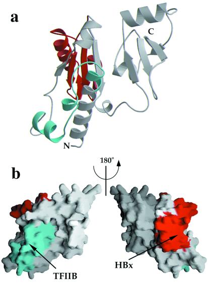
Figure 4.
Location of regions that have been proposed to interact with TFIIB and hepatitis B virus protein X (HBx). (a) Ribbon diagram representation of yRPB5 (in the same orientation as in Fig. 2). The fragment corresponding to the homologous sequence in hRPB5 that has been shown to interact with hTFIIB is shown in cyan whereas the region that is responsible for HBx interaction is shown in red. (b) Representation of the molecular surface of yRPB5 in the same orientation as Fig. 2 (Left) and rotated by 180° around the vertical axis (Right), showing the exposed surfaces of the above regions.

