
Figure 7. Pattern of mRNA expression of neurotrophic factors and myelin proteins in spinal cords of rats.
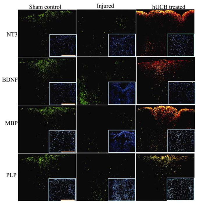
FISH analysis of neurotrophic factors and myelin proteins depicting sham control, injured, and hUCB-treated samples in the dorsal white matter region. Sequential serial sections hybridized with FITC-conjugated oligonucleotide antisense probes for NT3, BDNF, MBP and PLP were photographed using confocal microscope as described in Methods section. hUCB treated sections show colocalization (yellow) with Texas-red conjugated CD44 antibody, specific for hUCB. Inset shows representative Hoechst -33342 stained images. Scale Bar = 100 μm. Results are from three independent sections caudal from the injury epicenter (n ≥ 3).