
Figure 6.
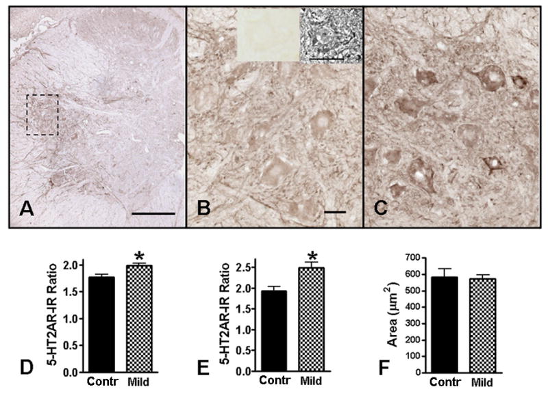
5-HT2A receptor immunoreactivity (5-HT2AR-IR) in the rostral dorsolateral nucleus (rDLn) of the L5-L6 spinal cord. B is a higher magnification photo of the enclosed area of the rDLn in A (uninjured control). The inserts on top of B are phase contrast (right) and brightfield (left) photos of a motoneuron in a section of the same uninjured spinal cord that has been processed without 5-HT2AR antibody (i.e., a no primary control). C is a representative higher magnification photo of the same region in a 4wk Mild SCI animal. In uninjured controls, the 5-HT2AR-IR was strongest around the peripheral rim and motoneuron pools in the ventral horn gray matter (A). At 4wks after Mild SCI (C), the 5-HT2AR-IR was increased compared to uninjured controls (B). Quantification of the optical density of 5-HT2AR-IR indicated a significant increase in the rDLn (D) as well as in the somas of the individual motoneurons located within the rDLn (E) of 4wk Mild SCI animals (n = 5) compared to uninjured controls (n = 4). The average motoneuron area in the uninjured controls and the 4wk Mild SCI animals was not statistically different from each other (F), thus did not contribute to the differences in 5-HT2AR-IR. Scale bar in A = 300μm. Scale bars in B, C = 30μm. *p<0.05 as compared to controls, unpaired t-test.