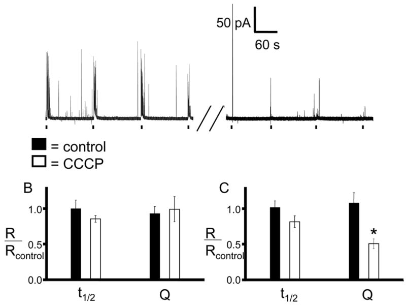
Figure 3. Amperometric detection of time-dependent depletion of epinephrine from vesicles.

Individual chromaffin cells were exposed 4 times to a 3-s pressure ejection of 60 mM K+in a Ca2+ containing buffer. The cells were then incubated in buffer containing 2 μM CCCP for the indicated time. After the incubation period, the cells were washed with buffer and stimulated again using the same K+ ejection pattern. Average spike parameters were then normalized by the pre-incubation values for each cell so that each cell acted as its own control. A) A representative amperometric trace of a 15 minute CCCP incubation. B) 5 minute incubation in CCCP caused no significant change in the average current spike half-width or quantal size compared with pre-incubation averages. C) 15 minute incubation causes a significant decrease in quantal size (p < 0.001 by Mann-Whitney U-test).
