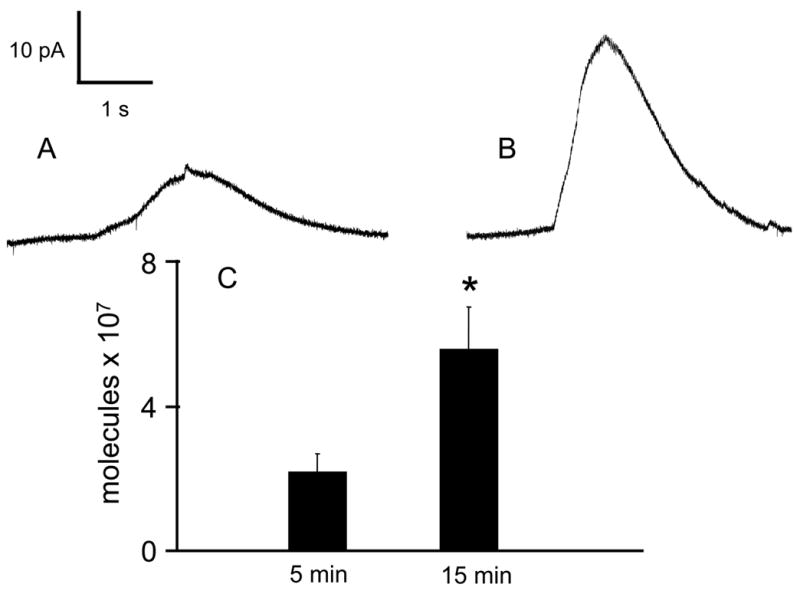
Figure 4. Cytosolic catecholamine content evaluated by digitonin permeabilization.

The cells were incubated with 2 μM CCCP in Ca2+-containing buffer for the either A) 5 minutes or B) 15 minutes. After washing and transferring to Ca2+-free buffer, 10 μM digitonin was pressure ejected onto the cell. The resulting envelope is an indication of the amount of catecholamine that was present in the cytosol of the cell. C) The 15 minute incubation resulted in approximately double the amount of catecholamine molecules that were detected after a 5 minute incubation (p < 0.05 by Mann-Whitney U-test).
