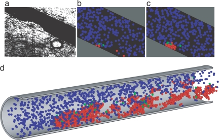
Fig. 1.
Evolution of thrombus growth. (a) Thrombus growing on a blood vessel wall in vivo is shown. [Reproduced with permission from ref. 1 (Copyright 1970, Nature Publishing Group).] (b) Similarly scaled computer model showing the early growth stage of thrombus is shown. Blue indicates unactivated platelets, green indicates triggered platelets, and conversion after a characteristic time delay to activated is indicated by red. (c) Later stage in thrombus growth at steady flow is shown. (d) Shown is a late stage in a similar computation, where adhesion of activated platelets is allowed at all locations downstream.

