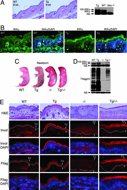
Fig. 2.
Overexpression of IKKα in the epidermis enhances terminal differentiation in the skin. (A) Tissue specimens of skin of WT and Lori·IKKα-Tg (Tg) mice stained with H&E. Transgenic IKKα expression (HA) in skin of WT and Tg mice was analyzed by Western blotting. IKKα, endogenous IKKα; HA-IKKα, transgenic IKKα; NS, nonspecific band; 8-wk, 8-week-old. (B) Elevated IKKα expression in the suprabasal epidermis in Tg mice by immunofluorescent staining. Green, IKKα staining; blue, DAPI nuclear staining; BL, basal layer; Ep, epidermis. (C) Appearance of WT, Tg, Ikkα−/− (−/−), and Lori·IKKα-Tg/Ikkαα−/− (Tg/−/−) newborn mice. (D) Filaggrin levels in WT, Tg, −/−, and Tg/−/− skin. NS, nonspecific band. (E) Comparison of involucrin (Invol) and filaggrin (Filag) expression in skin of WT, Tg, −/−, and Tg/−/− newborn mice, analyzed by immunofluorescent staining. Red, involucrin or filaggrin staining; blue, DAPI nuclear staining; Ep, epidermis between two arrows. White line separates epidermis and dermis.

