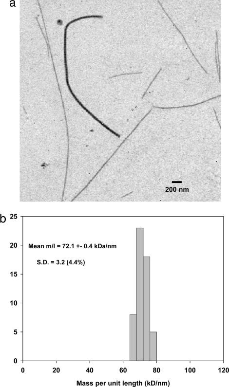
Fig. 1.
STEM data for collagen fibrils from 14-d chick embryonic sternum. (a) Annular dark-field STEM image of unstained fibrils released by mechanical disruption is shown. This represents a typical field of view showing several thin fibrils and a single thick fibril. The thick fibril has a measured M/L of ≈500 kDa/nm compared with ≈70 kDa/nm for the thin fibrils. (b) Histogram shows the M/L distribution of the thin fibril component measured from STEM images similar to that shown.

