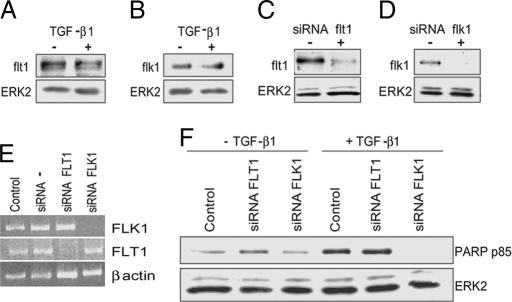
Fig. 4.
flk-1 mediates the apoptotic activity of TGF-β1. (A and B) Western blotting analysis of flt-1 and flk-1 expression in HUVEC incubated without (−) or with (+) TGF-β1 (1 ng/ml) for 6 h. ERK2: loading control. (C and D) Western blotting. (E) RT-PCR. Shown is flt-1 or flk-1 expression in HUVEC transfected with flt-1 or flk-1 siRNAs (+) or mock-transfected (−). ERK2 and β-actin: loading controls. Control, nontransfected cells; siRNA −, mock-transfected cells. (F) Western blotting analysis of PARP degradation in HUVEC transfected with flt-1 or flk-1 siRNAs or mock-transfected (Control). These experiments were repeated three times with similar results.

