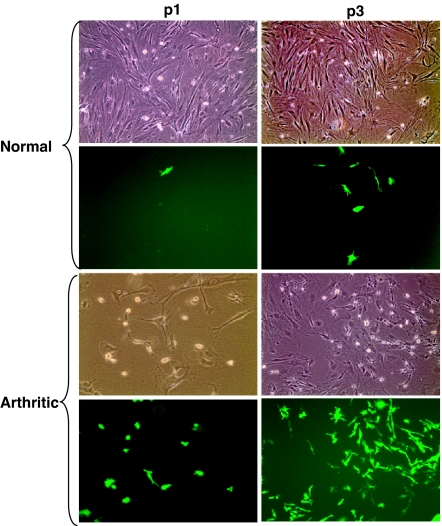
Fig. 1.
The presence of BM-derived cells in the populations of normal and arthritic FLS. BM cells from GFP transgenic donor mice were transferred into lethally irradiated GFP-negative recipient mice. Two months later, primary FLS were established by an enzymatic dispersal of synovial explants of the recipient BMT mice. p1 and p3, the first and third passages of primary FLS. The first and third rows represent phase-contrast microscopy; the second and fourth rows represent fluorescent microscopy. Normal, primary FLS that were obtained from normal joints of the BMT mice. At the third passage, GFP-positive cells comprised 1.2 ± 0.2% of the population of normal FLS (mean ± SEM of two experiments). Arthritic, primary FLS that were obtained from arthritic BMT recipient mice as described in Materials and Methods. At the third passage, GFP-positive cells comprised 33.7 ± 1.6% of the population of arthritic FLS (mean ± SEM of three experiments). (Original magnification: ×100.)

