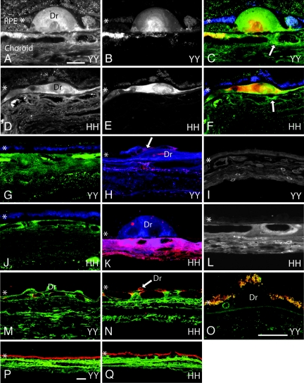
Fig. 1.
Confocal immunofluorescence images from CFH 402YY and 402HH donor eyes. (A–F) CFH immunoreactivity is similar within the choroid and in drusen (Dr) of 402YY [A and C (green)] and 402HH [D and F (green)] individuals. Arrows in C and F highlight focal regions of intense CFH immunoreactivity in the choroid adjacent to drusen. Focal C5b-9 immunoreactivity in drusen and in the choroid is also similar between 402YY [B and C (red)] and 402HH [E and F (red)] donor eyes. (G and J) CFH immunoreactivity in normal choroidal regions (not associated with drusen) is similar between 402YY [G (green)] and 402HH [J (green)] individuals. (H, I, K, and L) Antibodies to CRP only faintly label the choroid, drusen, and drusen vesicles in two different 402YY donor eyes [H (red) and I] but intensely label drusen vesicles, the extracellular matrix, endothelial cells, fibroblasts, and perivasculature regions of the choroid in two different 402HH donor eyes [K (red) and L]. RPE cells over drusen are only occasionally labeled with antibodies to CRP [H (arrow)] in both 402YY and 402HH genotypes. In the same sections, antibodies to CFH label the choroidal stroma and drusen [H and K (blue)]. (M, N, P, and Q) SAP immunoreactivity [M (402YY, green) and N (402HH, green)] and albumin immunoreactivity [P (402YY, green) and Q (402HH, green)] are similar in the choroid and in drusen of donor eyes with either CFH genotype. (O) Antibodies to the heparan sulfate proteoglycan perlecan only faintly label drusen vesicles and Bruch's membrane in tissue from both genotypes (402YY, green; 402HH, not shown). Lipofuscin autofluorescence demarcates the RPE [C, F, G, and J (blue); and M–Q (red/yellow)]. The RPE is identified with an asterisk (∗) in each panel. (Scale bars: A–N, 20 μm; O, 20 μm; P and Q, 20 μm.)

