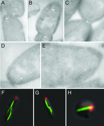
Fig. 4.
Polar localization of the MamK–GFP filament extremity. Localization of the MamK–GFP filaments in E. coli cells are revealed by immunogold staining (A–E, white arrows) or green fluorescence (F–H). Coexpressed IcsA–mCherry is shown by red fluorescence in the wild-type (F and G) and mreB mutant cell (H).

