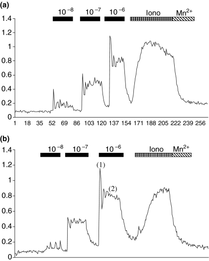
Figure 4.
Relative fluorescence against time (s). Cells from control (a) and 24-h ligated glands (b) were stimulated with increasing doses of methacholine (10−8–10−6m). Cells were then incubated with ionomycin (iono) and then manganese chloride (Mn2+) to elicit the maximum and minimum fluorescence signal. During stimulation by methacholine a biphasic response was seen comprising a peak (1) and plateau (2) phase.

