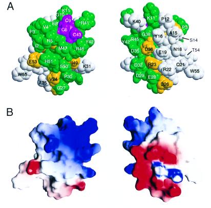
Figure 3.
Conserved residues and charged regions on the surface of mtRPB10. (A) Space filling model of mtRPB10 with conserved surface residues highlighted according to the color scheme of Fig. 1. Note the pronounced clustering of such residues around the zinc-binding site. (B) Surface charge distribution calculated and drawn by using grasp (34), with red indicating regions of negative potential and blue of positive potential. The basic region, formed by Arg5, Lys11, Lys31, Lys40, Arg41, Arg45, and Arg46, is centered near the N terminus and along helix 3. The acidic region is located predominantly in helix 2 and the preceding loop because of Glu19, Asp26, Glu28, Asp29, Asp32, Asp35, and Asp36. The extended neutral region includes polar and hydrophobic residues near the N terminus (Met1, Ile2, Pro3, Val4, Pro12), along helix 1 (Ser14, Ala15, Tyr16, Asn18, Tyr20, Gln21), flanking the loop between helices 1 and 2 (Val24, Ala25, Pro30), and within or immediately after helix 3 (Tyr42, Met47, Ser50, His51). In both A and B, the two views differ by a rotation of 180° around the vertical axis.

