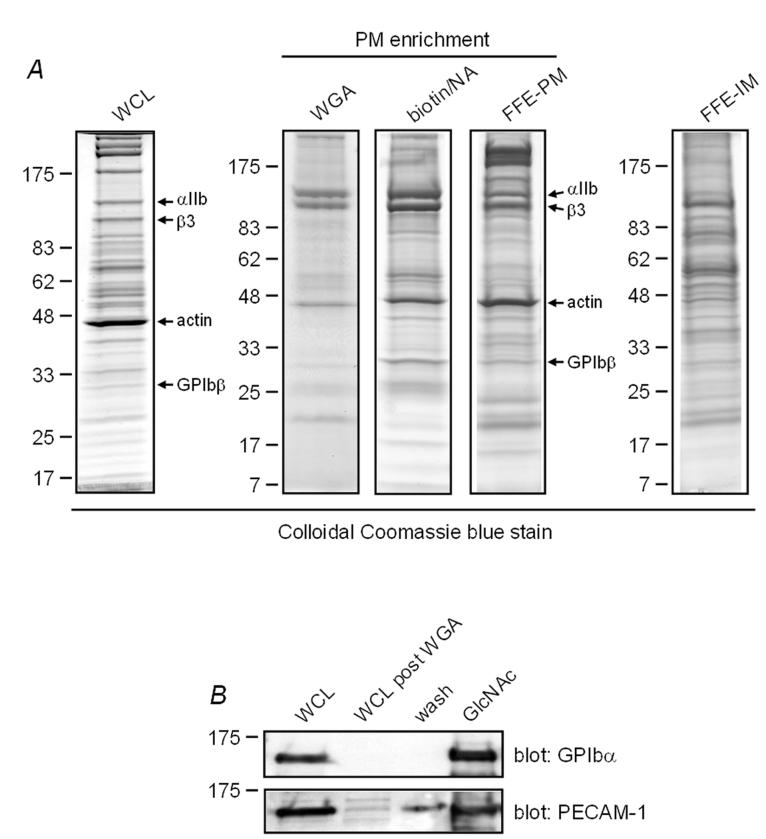
Fig. 1.
Comparison of proteins isolated by WGA affinity chromatography, biotin/NeutrAvidin (NA) affinity chromatography and free flow electrophoresis (FFE). A, platelet whole cell lysate (WCL) and proteins isolated by the three enrichment techniques were resolved on 4-20% SDS-PAGE gels and stained with Colloidal Coomassie blue. Bands corresponding to αIIb, β3, actin and GPIbβ were identified by tandem mass spectrometry and are shown to the left of the panels. WGA, wheat germ agglutinin affinity chromatography; biotin/NA, biotin/NeutrAvidin affinity chromatography; FFE-PM, free flow electrophoresis-plasma membrane fraction; FFE-IM, free flow electrophoresis-intracellular membrane fraction. Images shown are representative of three WGA, three biotin/NA, and two FFE enrichment experiments. B, aliquots taken at various stages of the WGA affinity chromatography procedure, including elution by N-acetylglucosamine (GlcNac), were western blotted for PECAM-1 and GPIbα.

