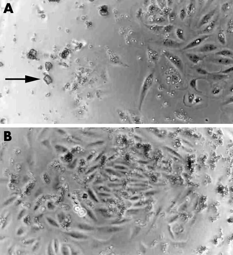
Figure 5 Triamcinolone acetonide, 0.10 mg/ml, in cell culture media. Phase microscopy 100× magnification (A). Retinal pigment epithelial cells are noticeably absent near the larger crystal clumps of the trade formulation (arrow). (B) A more uniform dispersion of triamcinolone crystals is noted in the preservative free formulation. Retinal pigment epithelial cells are distributed evenly and in the vicinity of the smaller crystals.
