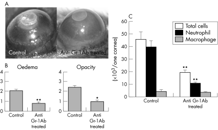
Figure 5 Corneal inflammation in mice depleted of neutrophils using anti‐Gr‐1 antibody. (A) Photographs of the cauterised corneas after 96 hours. (B) The clinical evaluation of corneal inflammation. C57/BL6 mice (n = 20), anti‐Gr‐1 antibody treated C57/BL6 mice (n = 20). *p<0.05; **p<0.01. (C) The analysis of corneal cell infiltrates 96 hours after cauterisation in untreated WT mice and WT mice treated with anti‐Gr‐1 antibody. **p<0.01.

An official website of the United States government
Here's how you know
Official websites use .gov
A
.gov website belongs to an official
government organization in the United States.
Secure .gov websites use HTTPS
A lock (
) or https:// means you've safely
connected to the .gov website. Share sensitive
information only on official, secure websites.
