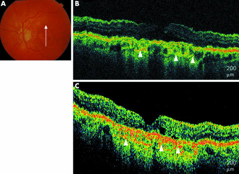
Figure 7 Geographic atrophy. (A) Colour fundus photograph of the left eye. (B) UHR‐OCT image of the left eye with generalised retinal thinning, hyporeflectivity of the RPE, and prominent choroidal vessels (white arrowheads). (C) StratusOCT image of the left eye shows a similar pattern in less detail (white arrowheads).

An official website of the United States government
Here's how you know
Official websites use .gov
A
.gov website belongs to an official
government organization in the United States.
Secure .gov websites use HTTPS
A lock (
) or https:// means you've safely
connected to the .gov website. Share sensitive
information only on official, secure websites.
