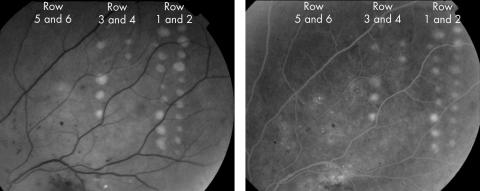
Figure 1 Retinal images taken immediately after diode laser photocoagulation of patient 2. (A) Red‐free fundus photograph. Two rows of continuous wave lesions are shown on the right. Micropulse lesions are shown in rows 3–6, as described in table 1. Haemorrhages and exudates were present preoperatively and are of diabetic origin. Only continuous wave and higher power micropulse lesions are visible. (B) A late phase fluorescein angiogram frame showing laser lesions and leakage from diabetic microaneurysms and telangiectatic vessels. All angiographically apparent lesions were also apparent on red‐free photographs.

An official website of the United States government
Here's how you know
Official websites use .gov
A
.gov website belongs to an official
government organization in the United States.
Secure .gov websites use HTTPS
A lock (
) or https:// means you've safely
connected to the .gov website. Share sensitive
information only on official, secure websites.
