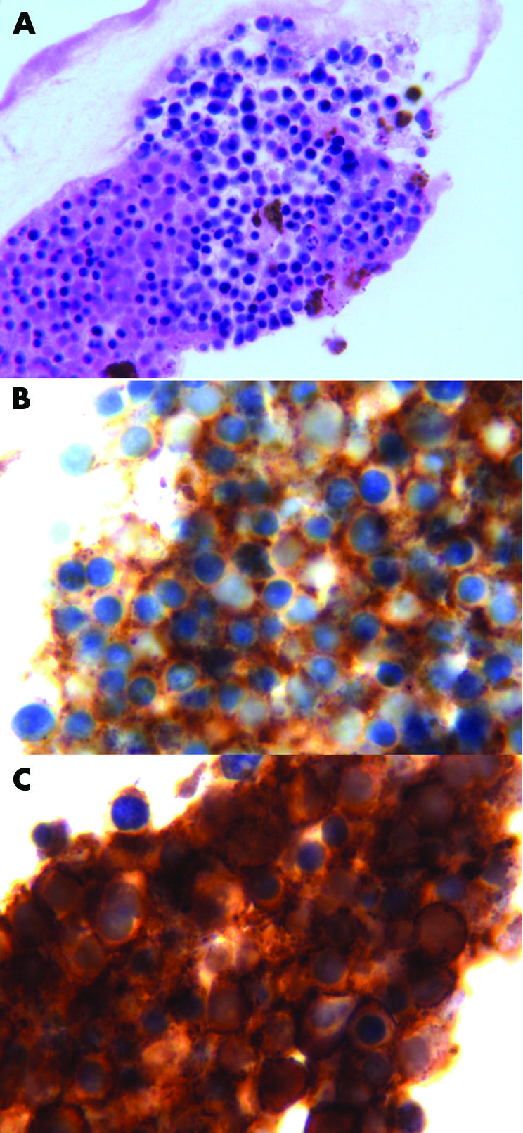
Figure 3 Case 3. (A) Cytological examination revealed numerous medium sized monotonous malignant lymphocytes with moderate pleomorphism and focal necrotic cells (haematoxylin and eosin staining; original magnification × 100). (B) Immunohistochemical staining demonstrated the cells were positive for CD 4 (CD 4 staining; original magnification × 100). (C) CD 30 positive (CD 30 staining; original magnification ×100).
