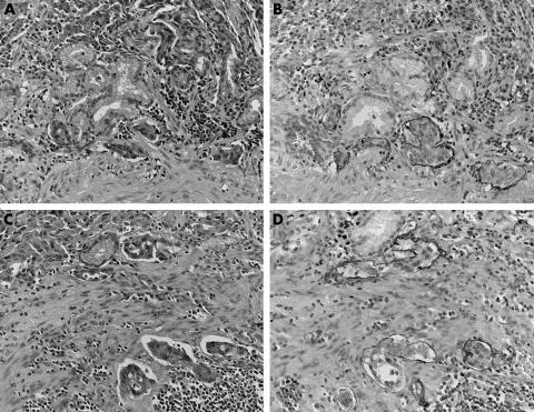
Figure 1 Immunohistochemical analysis for podoplanin in gastric cancer. Podoplanin positive lymph vessels can be seen from the muscularis mucosae to the superficial submucosa. (A–D) Invasion of the carcinoma cells into the lymph vessels was seen mostly in the muscularis mucosae and the submucosa. In some cases (A, B), lymphatic invasion was noted in the mucosa. Although it is difficult to identify lymph vessel invasion by haematoxylin and eosin staining (A, C), podoplanin immunohistochemistry made it easy to identify lymph vessel invasion (B, D).

An official website of the United States government
Here's how you know
Official websites use .gov
A
.gov website belongs to an official
government organization in the United States.
Secure .gov websites use HTTPS
A lock (
) or https:// means you've safely
connected to the .gov website. Share sensitive
information only on official, secure websites.
