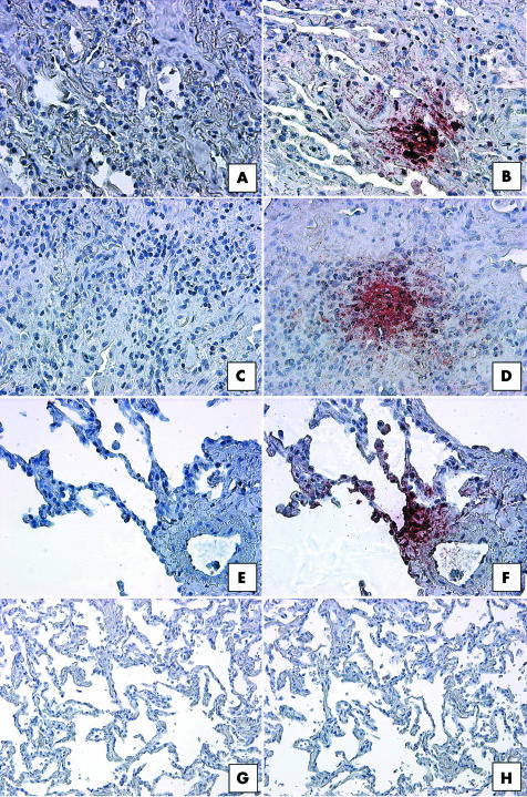
Figure 5 Representative immunohistochemical analysis of CCR7 in SLBs from (A, B) UIP, (C, D) NSIP, (E, F) RBILD, and (G, H) normal margin tumour patient groups. Panels (A), (C), (E), and (G) show control staining. CCR7 immunoreactivity (red staining) was seen in focal areas in SLBs from (B) UIP, (D) NSIP, and (F) RBILD, but not (H) normal margins. Original magnification, ×200. CCR7, CC chemokine receptor 7; NSIP, non‐specific interstitial pneumonia; RBILD, respiratory bronchiolitis/interstitial lung disease; SLB, surgical lung biopsy; UIP, usual interstitial pneumonia.

An official website of the United States government
Here's how you know
Official websites use .gov
A
.gov website belongs to an official
government organization in the United States.
Secure .gov websites use HTTPS
A lock (
) or https:// means you've safely
connected to the .gov website. Share sensitive
information only on official, secure websites.
