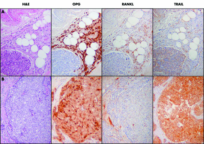
Figure 4 (A) A grade 2 lobular breast cancer. There is strong nuclear and cytoplasmic staining for OPG (osteoprotegerin), which contrasts with the negative normal lobule in the bottom left of the image. There is weaker focal staining for RANKL (receptor activator of nuclear factor κB ligand) and the tumour is negative for TRAIL (tumour necrosis factor related apoptosis inducing ligand). (B) A grade 3 ductal breast cancer of no specific type. There is strong nuclear and cytoplasmic staining for OPG, strong cytoplasmic staining for TRAIL, but the tumour is negative for RANKL. H&E, haematoxylin and eosin.

An official website of the United States government
Here's how you know
Official websites use .gov
A
.gov website belongs to an official
government organization in the United States.
Secure .gov websites use HTTPS
A lock (
) or https:// means you've safely
connected to the .gov website. Share sensitive
information only on official, secure websites.
