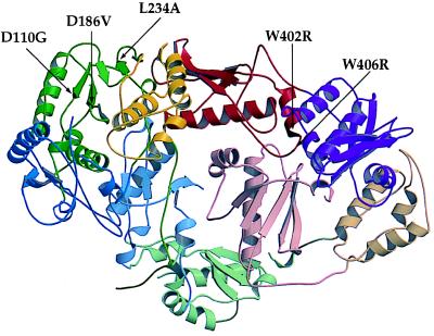
Figure 5.
Ribbon diagram of unliganded HIV-1 RT showing position of L234A primer grip mutation and locations of suppressors (shaded black). The figure was generated by molscript (38) and raster3d (39) with coordinates (2) retrieved from the Research Collaboratory for Structural Bioinformatics (RCSB) Protein Data Bank (PDB) (http://www.rcsb.org/pdb; PDB ID code 1HMV). Domains are defined as in ref. 3: fingers, blue; palm, green; thumb, yellow; connection, red; RNase H, purple. Domains in p66 are in fully saturated colors whereas in p51 they have decreased saturation. Secondary structure was assigned by using dssp (40). Spirals represent α-helices; arrows denote β-strands.

