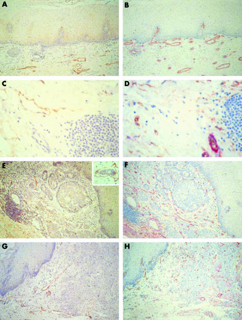
Figure 1 Representative immunohistochemical staining of normal oesophagus ((A, B); (C, D) submucosa) and adenocarcinoma ((E, F) differentiated and (G, H) undifferentiated) stained for (A, C, E, G) LYVE‐1 or (B, D, F, H) CD34 (brown) and α smooth muscle actin (red). Original magnification, ×200 (A, B, E–H); ×400 (C, D). Insert in (E), tumour cell invasion into peritumorous lymphatic vessel.

An official website of the United States government
Here's how you know
Official websites use .gov
A
.gov website belongs to an official
government organization in the United States.
Secure .gov websites use HTTPS
A lock (
) or https:// means you've safely
connected to the .gov website. Share sensitive
information only on official, secure websites.
