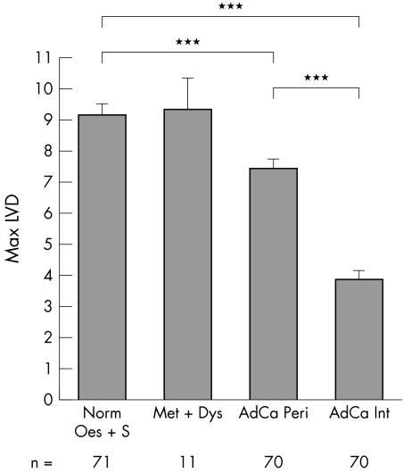
Figure 2 Maximum lymphatic vessel density (LVD) in the neoplastic progression: normal oesophagus and stomach (Norm Oes + S), metaplasia and dysplasia (Met + Dys), and peripheral or internal adenocarcinoma (AdCa Peri/AdCa Int); unpaired samples. ***p < 0.001); n, number of patients/group. Representative immunohistochemical images stained with LYVE‐1 are shown in fig 1A and C (normal oesophagus) and fig 1E and G (adenocarcinoma).

An official website of the United States government
Here's how you know
Official websites use .gov
A
.gov website belongs to an official
government organization in the United States.
Secure .gov websites use HTTPS
A lock (
) or https:// means you've safely
connected to the .gov website. Share sensitive
information only on official, secure websites.
