
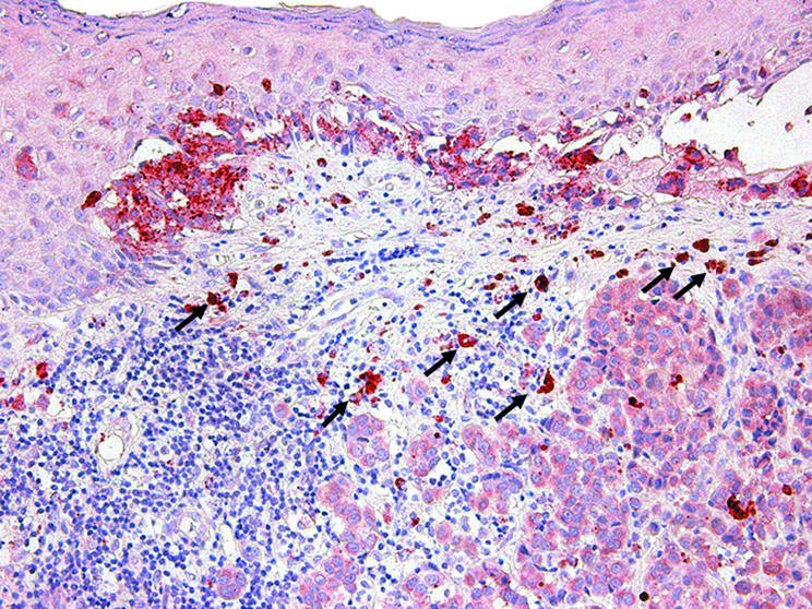
Figure 7 Immunohistochemical staining for vascular endothelial growth factor C (VEGF‐C) within the neoplastic cells of a thin cutaneous melanoma, both within the in situ and invasive components. Note that VEGF‐C expression is also detected within epidermal keratinocytes overlying melanoma cells, scattered dermal fibroblasts, and macrophages (arrows).