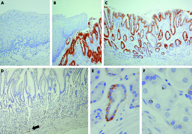
Figure 3 EpCAM immunohistochemistry performed on (A) normal oesophageal mucosa, (B–C) Barrett's metaplasia, and (D–F) normal gastric body mucosa. In particular, (B) demonstrates the sharp contrast in EpCAM expression shown by squamous and Barrett's epithelia. (D) Low power examination of gastric mucosa showed no obvious EpCAM immunostaining. (F) However, at higher power, scattered cells at the gland bases (the gland base shown in panel (E) is arrowed in panel (D)) were found to express EpCAM; these labelled cells had the morphological appearance of mucus secreting cells. (F) In contrast, neither chief (Ch) nor parietal (Pr) cells showed any immunostaining for EpCAM.

An official website of the United States government
Here's how you know
Official websites use .gov
A
.gov website belongs to an official
government organization in the United States.
Secure .gov websites use HTTPS
A lock (
) or https:// means you've safely
connected to the .gov website. Share sensitive
information only on official, secure websites.
