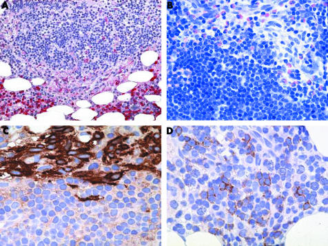
Figure 2 SM‐AHNMD (SM‐CLL) involving the bone marrow. (A) The naphthol AS‐D chloroacetate esterase (CAE) stain shows a mixed compact infiltrate consisting predominantly of small (CAE negative) lymphocytes with perifocally aggregated spindle shaped (moderately CAE positive) mast cells. Note the intact haematopoiesis in the lower part of the picture with prominent (strongly CAE positive) neutrophilic granulocytopoiesis. (B) Giemsa stain again demonstrates a relatively monomorphic lympoid infiltrate with an abundance of small lymphocytes. These cells contain only small amounts of cytoplasm and exhibit inconspicuous nucleoli. Blast cells are virtually absent. Note the atypical hypogranulated spindle shaped mast cells and the intermingled eosinophils in the upper right quadrant of the picture. (C) Anti‐tryptase antibody AA1 clearly decorates the compact cohesive nature of the mast cell infiltrate and demarcates it sharply from the surrounding lymphocytes. (D) A major proportion of lymphocytes expressed CD23, which can be regarded as clear indication of a neoplastic immunophenotype with a background of lymphocytic lymphoma. Original magnification: (A)×25; (B)×100; (C, D)×200.

An official website of the United States government
Here's how you know
Official websites use .gov
A
.gov website belongs to an official
government organization in the United States.
Secure .gov websites use HTTPS
A lock (
) or https:// means you've safely
connected to the .gov website. Share sensitive
information only on official, secure websites.
