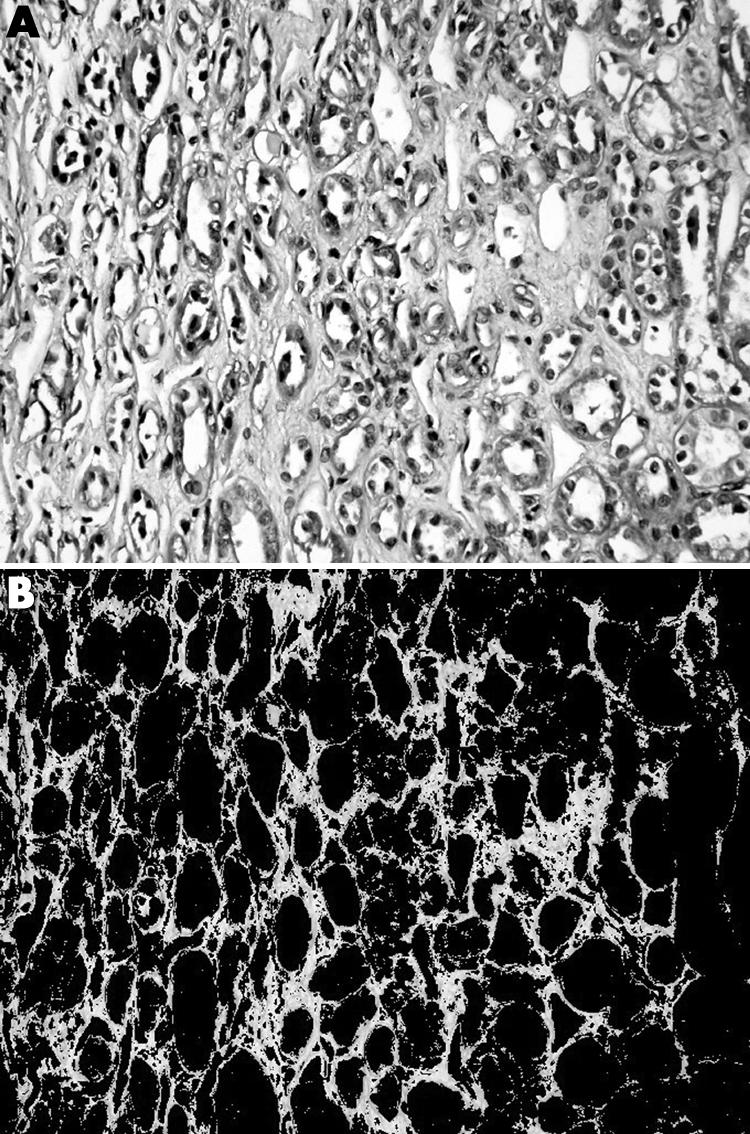
Figure 2 Masson trichrome stained section of a renal allograft biopsy demonstrating interstitial fibrosis (A) before and (B) after the selection for image analysis measurement of the green stained area.

Figure 2 Masson trichrome stained section of a renal allograft biopsy demonstrating interstitial fibrosis (A) before and (B) after the selection for image analysis measurement of the green stained area.