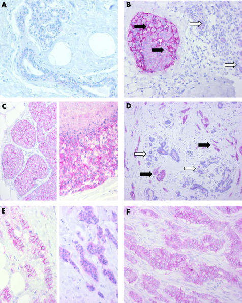
Figure 1 Activated leucocyte cell adhesion molecule (ALCAM) immunohistochemistry of breast glands and breast cancer. (A) Normal breast duct and gland epithelium with minimal ALCAM expression. (B) Ductal carcinoma in situ (black arrows) with strong membranous and moderate cytoplasmic staining; normal breast glands (white arrows) with weak cytoplasmic staining. (C) Lobular carcinoma in situ (on the left) with strong membranous and weak to moderate cytoplasmic staining; high grade ductal carcinoma in situ (on the right) with comedo necrosis and strong membranous and cytoplasmic staining. (D) Invasive ductal carcinoma (black arrows) with strong membranous and cytoplasmic staining; normal breast glands with weak cytoplasmic staining (white arrows). (E) Invasive ductal carcinoma with strong membranous and weak cytoplasmic staining (on the left) and a different case with strong cytoplasmic staining lacking membranous immunoreactivity (on the right). (F) Invasive ductal carcinoma with strong membranous and moderate cytoplasmic staining.

An official website of the United States government
Here's how you know
Official websites use .gov
A
.gov website belongs to an official
government organization in the United States.
Secure .gov websites use HTTPS
A lock (
) or https:// means you've safely
connected to the .gov website. Share sensitive
information only on official, secure websites.
