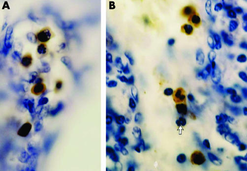
Figure 3 (A) Focus of immunostained fetal haemoglobin‐normoblasts (FNBS) in adenocarcimona of the colon. The three upper FNBS are in mitosis. (B) Immunostained fetal haemoglobin‐myeloid cells (FMLC) as segmented (middle) and juvenile (above and below) cells in a local lymph node of infiltrating ductal carcinoma of the breast. The arrow indicates a normal, unstained segmented myeloid cell.

An official website of the United States government
Here's how you know
Official websites use .gov
A
.gov website belongs to an official
government organization in the United States.
Secure .gov websites use HTTPS
A lock (
) or https:// means you've safely
connected to the .gov website. Share sensitive
information only on official, secure websites.
