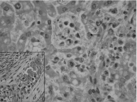
Figure 2 High‐power photomicrograph showing malignant histiocytes infiltrating and distending the sinusoids of the liver. Cytophagocytosis by a tumour cell can be seen just above the centre of the field (arrow). The inset shows blood vessel permeation in the myometrium by tumour cells (haematoxylin–eosin staining, 400×).

An official website of the United States government
Here's how you know
Official websites use .gov
A
.gov website belongs to an official
government organization in the United States.
Secure .gov websites use HTTPS
A lock (
) or https:// means you've safely
connected to the .gov website. Share sensitive
information only on official, secure websites.
