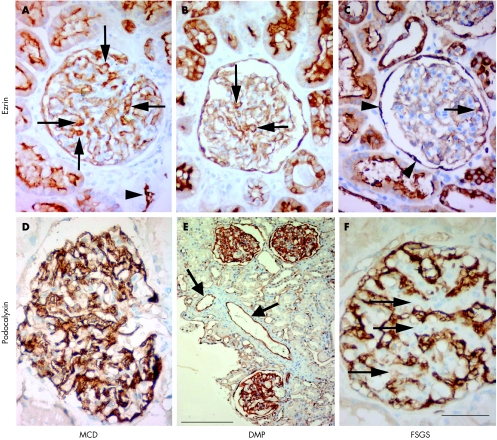
Figure 1 Light microscopy of renal glomeruli in children with immunohistochemical reactions for ezrin (A–C) and podocalyxin (D–F). Scale bar: 50 μm (A–D,F), 200 µm (E). (A) Immunohistochemical expression of ezrin in podocytes (arrows), parietal glomerular epithelial cells (Bowman's capsule) and on the apical surface of tubular epithelial cells (arrowhead) in a10‐year‐old child with minimal change disease (MCD). (B) Renal glomerulus of a 9‐year‐old child with diffuse mesangial proliferation (DMP). Ezrin immunoreactivity in podocytes (arrows) and tubular epithelial cells. Note the decreased number of ezrin‐positive podocytes and the lower staining intensity compared to A. (C) Glomerulus of an 11‐year‐old child with focal segmental glomerulosclerosis (FSGS) with only individual ezrin‐positive podocytes (arrow). Parietal glomerular epithelial cells (arrowheads) and tubular epithelial cells show positive reactions for ezrin. (D) Intensive immunohistochemical reaction for podocalyxin in the glomeruli (10‐year‐old child with MCD). (E) Renal cortex of an 11‐year‐old child with DMP and steroid‐resistant idiopathic nephrotic syndrome. Some podocytes with a positive reaction for podocalyxin. The arrows show extra‐glomerular endothelial cells with a positive reaction for podocalyxin. (F) Renal glomerulus of a 9‐year‐old child with FSGS. There is no reaction for podocalyxin in areas of sclerosis (arrows).

An official website of the United States government
Here's how you know
Official websites use .gov
A
.gov website belongs to an official
government organization in the United States.
Secure .gov websites use HTTPS
A lock (
) or https:// means you've safely
connected to the .gov website. Share sensitive
information only on official, secure websites.
