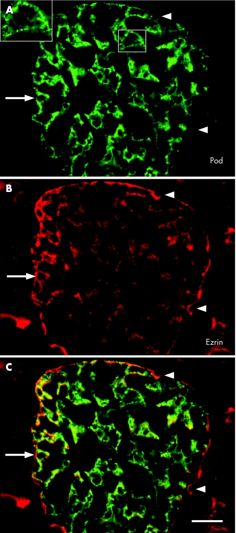
Figure 3 (A–C) Podocalyxin (pod) and ezrin in a renal glomerulus of a 10‐year‐old child presenting with diffuse mesengial proliferation. The large inset (magnified from the small inset) shows the granular pattern of podocalyxin distribution. Note the largely overlapping immunofluorescence of both antibodies in podocytes (arrow; yellow in C). In addition, ezrin is located on the apical cell membranes (probably microvilli) of proximal tubuli and in parietal glomerular cells (Bowman's capsule; arrowhead in (B)). (C) Scale bar 20 µm.

An official website of the United States government
Here's how you know
Official websites use .gov
A
.gov website belongs to an official
government organization in the United States.
Secure .gov websites use HTTPS
A lock (
) or https:// means you've safely
connected to the .gov website. Share sensitive
information only on official, secure websites.
