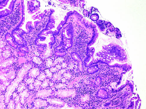
Figure 3 Focal gastric metaplasia. Enterocytes are replaced by foci of gastric‐type mucus‐secreting cells. Villi are shortened and blunted. The lamina propria shows an increase in the inflammatory cells (lymphocytes and plasma cells), associated with the hypertrophy and prolapse of Brunner's glands. These features are typical of non‐specific chronic duodenitis (haematoxylin–eosin staining, ×100).

An official website of the United States government
Here's how you know
Official websites use .gov
A
.gov website belongs to an official
government organization in the United States.
Secure .gov websites use HTTPS
A lock (
) or https:// means you've safely
connected to the .gov website. Share sensitive
information only on official, secure websites.
