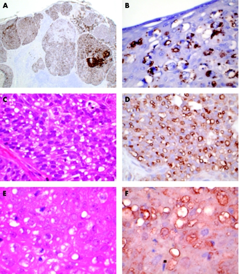
Figure 2 Immunohistochemical staining for adipophilin, perilipin and TIP47/PP17 in sebaceous gland carcinoma (SGC). (A) Immunohistochemical staining for adipophilin of SGC shown in fig 1B. There is widespread strong positivity, with numerous cytoplasmic vacuoles (anti‐adipophilin, ×100). (B) Immunohistochemical staining for adipophilin of SGC shown in fig 1D. There is strong positivity staining of vacuoles highlighting the Pagetoid infiltration of the epidermis (anti‐adipophilin, ×400). (C) Moderately differentiated nodular SGC showing numerous intracytoplasmic vacuoles (haematoxylin and eosin (H&E), ×400). (D) Immunohistochemical staining for perilipin outlines numerous small lipid vacuoles within the tumour cells (anti‐perilipin, ×400). (E) Moderately differentiated nodular SGC showing numerous intracytoplasmic vacuoles (H&E, ×400). (F) Immunohistochemical staining for TIP47/PP17 outlines numerous small lipid vacuoles in the tumour cells (anti‐TIP47/PP17, ×400).

An official website of the United States government
Here's how you know
Official websites use .gov
A
.gov website belongs to an official
government organization in the United States.
Secure .gov websites use HTTPS
A lock (
) or https:// means you've safely
connected to the .gov website. Share sensitive
information only on official, secure websites.
