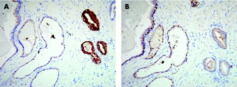
Figure 4 Prostatic adenocarcinoma showing cytoplasmic staining in the neoplastic glands for P504S (α‐methylacyl‐CoA racemase) in contrast with the negative adjacent benign atrophic glands (A), which show a preserved basal cell layer with nuclear staining for p63 in the consecutive section (B). Basal cells are absent in the malignant glands.

An official website of the United States government
Here's how you know
Official websites use .gov
A
.gov website belongs to an official
government organization in the United States.
Secure .gov websites use HTTPS
A lock (
) or https:// means you've safely
connected to the .gov website. Share sensitive
information only on official, secure websites.
